|
FOCUS-SIS (updated 02/04)
FOCUS-SIS stands for FOCUS-Subtracted Ictal-interictal Spect. This is a little program that helps locating epileptogenic foci in refractory partial epilepsy, thanks to nuclear medicine images.
I wrote FOCUS-SIS in Java as an ImageJ plugin.
ImageJ
ImageJ is a public domain Java Image processing program that can display, edit, analyze and process images. The source code is freely available and plugins can be built and added easily thanks to a powerful API.
ImageJ and some of its plugins were written by Wayne Rasband, who is at the Research Service Branch, National Institute of Mental Health, Bethesda, Maryland, USA.
ImageJ can be downloaded at http://rsb.info.nih.gov/ij.
A large amount of plugins can be found at http://rsb.info.nih.gov/ij/plugins.
A very useful introduction to writing plugins can be found here : http://mtd.fh-hagenberg.at/depot/imaging/imagej/.
How FOCUS-SIS does work ?
Download (http://rsb.info.nih.gov/ij) and install ImageJ.
Download the zip file FOCUS-SIS.zip, unzip it in the plugins folder of ImageJ. This latter file contains the source code (.java) and executable (.class), as well as two example images that can be read with the Analyze_Reader plugin
(http://rsb.info.nih.gov/ij/plugins/analyze.html).
Reload ImageJ; FOCUS-SIS now appears in the Plugins menu.
This plugin normalizes and subtracts two SPECT (nuclear medicine) images to help neurologists locating epileptogenic foci in partial seizures.
A peri-ictal and an inter-ictal exams are needed; the are supposed to be spatially co-registered. A Graphical User Interface allows the user to choose between relative or absolute subtraction (Lassen's correction).
The normalization method used was awarded by the Alavi-Mandell prize and is described in : Boussion N, Houzard C, Ostrowsky K, Ryvlin P, Mauguière F, Cinotti L. Automated detection of local normalization areas for ictal-interictal subtraction brain SPECT imaging. J Nucl Med 2002; 43: 1419-1425.
(Please cite this reference for any publication or communication concerning the use of FOCUS-SIS !)
New : this version now supports 8, 16 and 32 bits images.
Very important : the plugin is freely delivered, with absolutely no warranty. It has been developped as a computer-aided decision tool, and should be used for this purpose only.
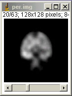
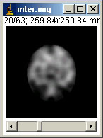
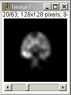
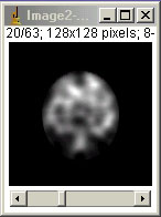
Two images (stacks of same size) must be open before starting FOCUS-SIS. For instance those in the zip file, per and inter :
When done, FOCUS-SIS can be launched, and a window appears :
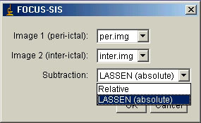
You can select Relative subtraction (percentage of signal increase), or absolute subtraction. This latter is performed by using Lassen correction (according to LASSEN NA et al. The retention of 99mTc-HMPAO in the human brain after intracarotid bolus injection: a kinetic analysis. J Cereb Blood Flow Metab 1988; 8(6): S13-22 and FRIBERG L et al. Retention of 99mTc-bicisate in the human brain after intracarotid injection. J Cereb Blood Flow Metab 1994; 14 Suppl 1:S19-27.).
If you select Lassen, a new window appears :
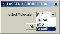
You have to input the name of the injected molecule, either HMPAO or ECD. If you do not know, select Default.
Then the normalization and the subtraction are performed. Three windows appear. The input images now normalized (they may require brightness/contrast
adjustment):
and the subtraction of these two latter, with the "fire" LUT :
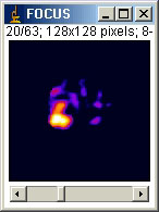
All these images are set to 32-bits.
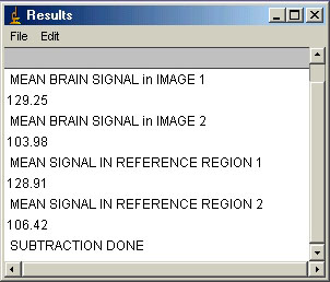
The Result window is also updated, showing the mean signal in the brain and in the reference region (when Lassen is selected) :
|